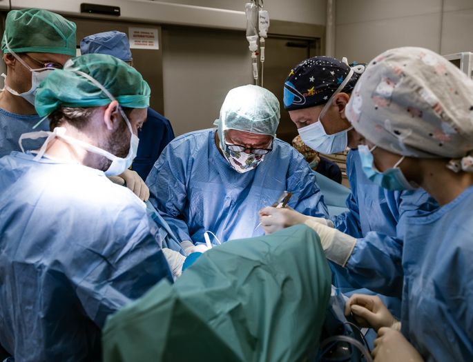
Complicated operations on internal organs will soon become much easier thanks to the technology developed by Jan Witowski, a student of the Jagiellonian University Medical College.
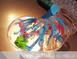
At the first stage of his work, Jan Witowski focused on creating and printing the model of a liver – the gland which plays a key role in the functioning of human organism by filtering blood, removing toxins, participating in the synthesis of cholesterol and triglycerides, converting carbohydrates into easily absorbable glucose, to mention only some of its important tasks.
The JU student aimed to create a technology which would make it possible to print a model of liver of a specific patient and is so cheap that it can be commonly used. Jan Witowski has carried out his research in the 2nd Chair of Surgery at the JU Medical College, under the supervision of Dr hab. Michał Pędziwiatr.
According to the article in Gazeta Prawna daily, “plastic parts of the model (reflecting the inside of the liver) are printed separately, and then silicon is poured over them and assumes the shape of a liver. The Polish scientist managed to find a material which is transparent (as the model needs to be) and can substitute transparent resin in expensive printers. Such a design of the process significantly contributes to the lowering of the printing cost. “Liver is an anatomically complicated organ and using our technology it can be printed for € 100-120, whereas in the case of other, foreign models the cost is over 2.5 thousand euro,” says Mr Witowski in the aforementioned article.
We asked Jan Witowski several questions about his work and plans for the future.
Piotr Żabicki, www.nauka.uj.edu.pl: What has to be done before pressing the “Print” button? What kind of information and parameters need to be determined?
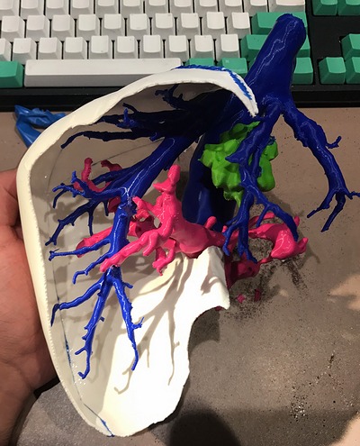
Jan Witowski: The models are primarily based on images, mainly obtained by means of computed tomography or magnetic resonance. It’s necessary to analyse where the anatomic structures that are interesting to us are, in order to single them out from the whole series of images. The process of singling out is called segmentation. It’s performed by means of proper computer algorithms, which make it possible to perform the operation relatively fast.
What are the specific uses of such printouts?
The models can be used for multiple purposes: for instance, we use liver models to plan surgical operations, but also to educate patients or students. This allows modernisation and personalisation of the entire treatment process, at each of its levels. Besides, the proper choice of materials could make it possible to create realistic simulators.
The technology you‘ve worked out is cheaper than its foreign counterparts. Does it mean that every hospital will be able to create such models?
This is really the goal, especially due to the fact that the a hospital-based “3D print shop” significantly shortens the time and lowers the cost of producing the models. At the largest medical centres, such as the Mayo Clinic or Stanford University, teams specially trained for that purpose have been existing for several years and the demand for such models is constantly increasing. In Poland this is a new area and the research group formed at the JU MC 2nd Chair of Surgery is the first such initiative in Poland.
What are the further plans regarding the development of this project?
At the moment, we’re conducting research that will assess the clinical importance of applying our models. Besides, we’ve started cooperation with other hospital units in Kraków and for several weeks have been printing models of aneurysms and hearts. Foreign medical centres have also showed their interest in such collaboration: for instance, we’re in contact with surgeons from Australia and the USA. Let me also add that our plans aren’t limited to 3D printing – we’ll test how other state-of-the-art technologies can be used to improve the treatment process and change patients’ life’s for the better.
--------------------------------------------------
Jan Witowski is a 4th year student of Medicine at the JU Medical College. Under the supervision of Dr hab. Michał Pędziwiatr, he conducts research on the medical application of 3D printing. His papers on technology in medicine have been published in prestigious scientific journals. He has given successful presentations both in Poland (Kraków, Wrocław, Lublin) and abroad (Stanford, Orlando).
Original text at: www.nauka.uj.edu.pl